Blood in the stool, a lasting bowel change, iron-deficiency anaemia, abdominal pain, or unexplained weight loss needs medical assessment rather than watchful waiting. By the end, you will understand which tests confirm colon cancer, how doctors determine its stage, and why treatment ranges from endoscopic removal to surgery, chemotherapy, targeted therapy, or immunotherapy.
Key takeaways
- Report blood in stool, persistent bowel changes, or unexplained weight loss for assessment.
- Screening can find precancerous polyps before they become colon cancer.
- Pathology and scans establish the cancer’s type, stage, and spread.
- Stage IV treatment depends on where the cancer has spread and its molecular findings.
What colon cancer is and when symptoms need assessment
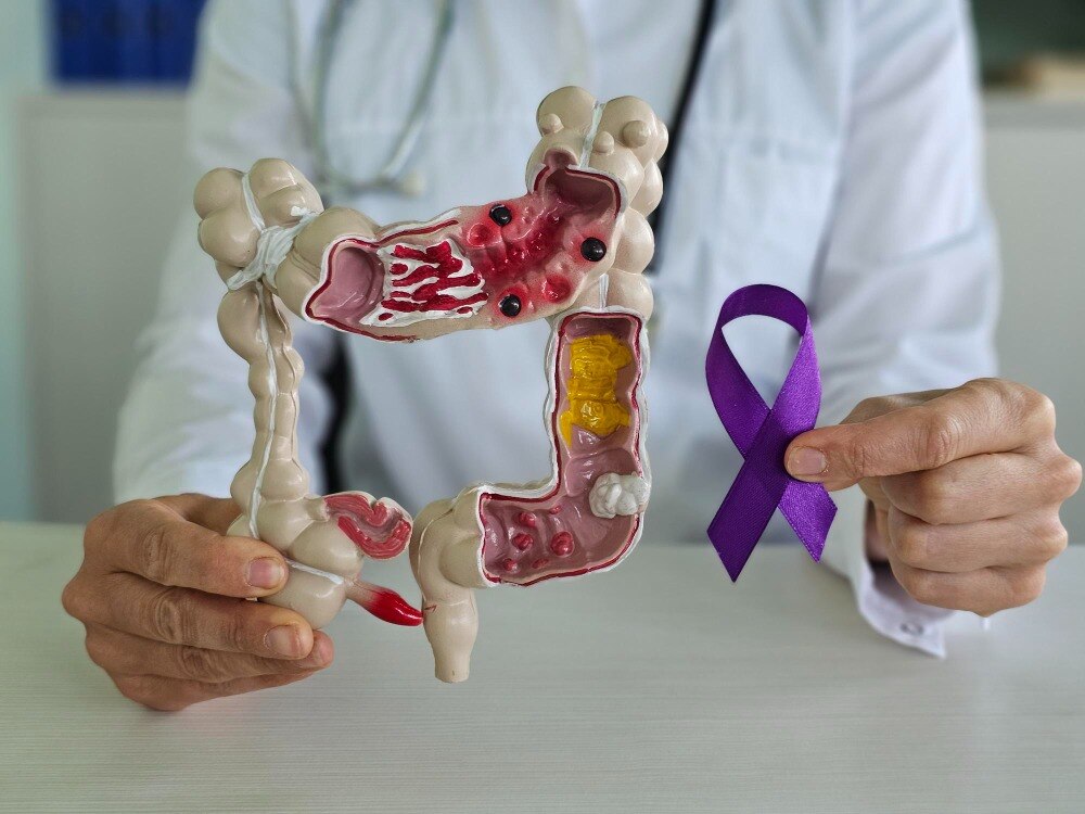
Colon cancer starts in the inner lining of the colon, the longest part of the large intestine, after the small intestine and before the rectum. The colon absorbs water and forms stool. Cancer in the rectum is rectal cancer, not colon cancer; both are colorectal cancers, but their treatment plans can differ.
Arrange medical assessment if you notice any of these colon cancer symptoms, especially if they persist, recur, or are worsening:
- Blood in stool, whether bright red or dark and tar-like
- A persistent change in bowel habits, including new constipation, diarrhoea, narrower stools, or urgency
- Unexplained iron-deficiency anaemia, fatigue, or breathlessness
- Ongoing abdominal pain, cramping, or bloating
- Unintentional weight loss or reduced appetite
These signs do not prove cancer, but they need an explanation. Seek prompt care for heavy rectal bleeding, black stools, severe pain, vomiting, or inability to pass stool or gas.
If you have no symptoms, screening still matters: average-risk adults should begin at age 45. Options include annual FIT, stool DNA-FIT every one to three years depending on the product, or colonoscopy every 10 years. People with family history, inflammatory bowel disease, or inherited syndromes need an individualized earlier plan.
A positive stool test requires diagnostic colonoscopy, not reassurance.
How screening and diagnostic testing find colon cancer
At the first appointment, the clinician separates screening from diagnostic testing. For average-risk adults, colon cancer screening commonly begins at age 45 with annual FIT or colonoscopy every 10 years. Symptoms, iron-deficiency anaemia, family history, inflammatory bowel disease, or an inherited syndrome require a diagnostic plan.
- The clinician reviews your symptoms, medicines, family history, and previous polyps, then examines your abdomen and rectum when appropriate.
- Blood tests can include a complete blood count, which may show anaemia. A normal result does not exclude cancer.
- A FIT test detects hidden blood in stool. A positive result is not a cancer diagnosis; it requires a diagnostic colonoscopy to find and remove polyps or sample a lesion.
- Before colonoscopy, you follow bowel-preparation instructions so the lining is visible. The endoscopist examines the entire colon, removes polyps, and takes tissue from suspicious areas.
A colonoscopy can show a tumour, but pathology establishes the diagnosis. The colon cancer biopsy is examined for malignant cells and tumour features that guide later treatment. CT scans of the chest, abdomen, and pelvis assess spread and surgical anatomy; they do not replace tissue diagnosis.
Dr.Parag Ingle - Surgical Oncologist can help interpret a positive FIT test or suspicious colonoscopy finding and coordinate the next diagnostic step, rather than treating a screening result as proof of cancer.
Related services
Colon cancer treatment Our Colon Cancer Treatment services aim to provide comprehensive care for individuals facing this health challenge. View service → |
Cancer is a disease that causes abnormal growth of cells in the body. It can affect any part of the body and can spread to other organs. View service → |
What pathology, scans, and staging reveal
A biopsy or resection specimen confirms colon cancer. CT scans cannot replace tissue diagnosis in the usual work-up.
The colon cancer pathology report identifies the tumour type, grade, depth of invasion, lymphovascular or perineural invasion, and whether the cut edge is clear. A resection specimen also shows how many regional lymph nodes contain cancer. Testing for mismatch repair proteins or microsatellite instability can influence treatment and inherited-cancer assessment.
Initial staging commonly includes a contrast-enhanced CT chest abdomen pelvis, a complete colon evaluation when feasible, and a baseline CEA test. CEA helps track a tumour that produces CEA, but a normal result does not exclude cancer, and an elevated result alone does not prove recurrence.
Doctors combine these findings using TNM staging: T describes invasion through the bowel wall, N describes involved regional nodes, and M describes distant spread.
- Stage 0: Cancer remains in the mucosa. Complete removal of a favourable malignant polyp can be curative.
- Stage I: Cancer has invaded the bowel wall but has not reached lymph nodes or distant organs.
- Stage II: Cancer has grown through or beyond the bowel wall without regional-node or distant spread.
- Stage III: Regional lymph nodes contain cancer, but there is no distant metastasis.
- Stage IV: Cancer has spread to distant organs, such as the liver or lungs; selected limited metastases can still be treated aggressively.
How treatment changes from stage 0 to stage IV
Stage determines the starting point, but tumour biology and whether every visible deposit can be removed often change the plan.
| Stage | Usual treatment | Features that change the plan |
|---|---|---|
| Stage 0 | Complete endoscopic removal; surgery if needed | Deep invasion, poor differentiation, lymphovascular invasion, or an involved or uncertain margin may require bowel surgery. |
| Stage I | Surgical removal of the affected colon segment and nearby lymph nodes | Complete removal with negative nodes usually means no chemotherapy. |
| Stage II | Surgery; stage II colon cancer chemotherapy is considered selectively | T4 invasion, obstruction, perforation, inadequate lymph-node sampling, lymphovascular invasion, or poor differentiation raises recurrence risk. MSI-high or deficient-MMR tumours usually gain little from fluoropyrimidine alone. |
| Stage III | Stage III colon cancer treatment is surgery followed by FOLFOX or CAPOX | Lower-risk tumours may receive three months of CAPOX to reduce cumulative nerve damage; higher-risk disease often needs longer treatment. |
| Stage IV | Systemic therapy, with surgery or ablation for selected metastases | Stage IV colon cancer treatment depends on RAS, BRAF V600E, MSI/MMR, HER2, and other results. Limited liver or lung spread may be resected or ablated with curative intent. |
A malignant polyp can be cured by endoscopic removal when the entire lesion is removed and pathology shows favourable features. An incomplete removal or high-risk pathology shifts treatment toward surgery.
For unresectable spread, molecular results guide drug selection: MSI-high or deficient-MMR disease can respond to checkpoint inhibitors, while RAS mutations rule out benefit from anti-EGFR antibodies. A multidisciplinary team reviews the primary tumour, metastases, pathology, scans, and your overall fitness before choosing treatment.
What happens after treatment and during surveillance
Doctors monitor for recurrence and new polyps through planned visits, blood tests, imaging, and colonoscopy. The schedule changes with stage, treatment, findings at the complete colonoscopy, and your fitness for further testing.
- Clinical reviews: Your doctor asks about new symptoms, examines you, and checks recovery at scheduled visits.
- CEA surveillance: If the original tumour produced carcinoembryonic antigen, blood tests can track it. Many plans measure CEA every 3–6 months for 2 years, then every 6 months through year 5. A normal result does not rule out recurrence, and one high result does not prove it.
- CT imaging: Contrast-enhanced CT of the chest, abdomen, and pelvis is scheduled for patients with meaningful recurrence risk. It can show suspicious disease but cannot replace tissue diagnosis.
- Surveillance colonoscopy: A complete examination checks the join or treated area and the remaining colon for new polyps or cancer. Many patients have one about 1 year after surgery; later examinations depend on what it finds. Large or numerous polyps usually lead to a shorter interval.
This planned colon cancer follow-up is only part of colon cancer recurrence monitoring. Contact your treatment team between appointments if a new symptom persists or worsens, rather than waiting for the next scheduled review.
Frequently asked questions
What symptoms of colon cancer need medical assessment?
Seek assessment for blood in stool, persistent changes in bowel habits, ongoing abdominal pain, iron-deficiency anaemia, or unexplained weight loss. These symptoms can have causes other than cancer, but they need evaluation.
How is colon cancer detected?
Screening may use stool tests or colonoscopy. If a colonoscopy finds a suspicious growth, a biopsy is examined by a pathologist to confirm cancer.
What do pathology, scans, and staging show?
Pathology identifies the cancer type and features in the tissue. CT or MRI scans show the tumour’s local extent and possible spread. Staging combines these findings to classify disease from stage 0 to stage IV.
How does treatment change from stage 0 to stage IV?
Stage 0 is usually treated by removing the abnormal area during colonoscopy or surgery. Stages I to III usually require surgery, with chemotherapy added for selected stage III and high-risk stage II cancers. Stage IV treatment focuses on systemic therapy, with surgery or local treatments for selected patients.
What happens after colon cancer treatment?
Follow-up commonly includes scheduled consultations, colonoscopy, blood tests such as CEA when appropriate, and scans when indicated. Your team sets the timing according to the original stage, treatment, recurrence risk, and symptoms.
Related service
Staging of cancer Cancer Staging is a crucial medical service that helps determine the extent and severity of cancer in a patient. View service → |