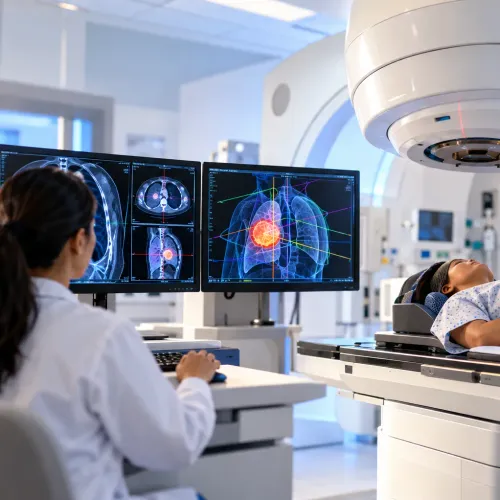
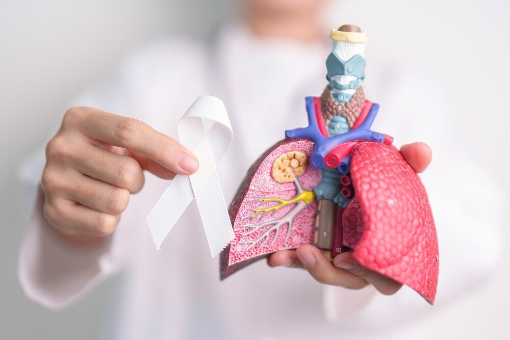
A cough, breathlessness or chest pain does not prove lung cancer, but symptoms that persist, worsen or recur deserve medical assessment because early lung cancer can remain silent. You will learn which warning signs and exposures matter, who may qualify for low-dose CT screening, how doctors confirm a diagnosis, and how treatment choices depend on cancer type, stage and your overall health.
Key takeaways
- Arrange assessment for a cough lasting more than three weeks.
- Seek prompt care for coughing blood, chest pain, or worsening breathlessness.
- Discuss low-dose CT screening if you have a heavy smoking history.
- Treatment depends on cancer stage, type, biomarkers, and your overall health.
Which symptoms need medical assessment?
Arrange medical assessment for lung cancer symptoms that persist, worsen, recur, or lack a clear explanation. Seek prompt care for a cough lasting more than three weeks, coughing blood, breathlessness and chest pain. Also report recurrent chest infections, unexplained weight loss, persistent fatigue, hoarseness, loss of appetite, or new finger clubbing.
Severe breathlessness or significant bleeding needs urgent attention.
Early lung cancer can cause no symptoms because a small tumour may not obstruct an airway or affect nearby tissues. Feeling well does not rule it out, especially in someone with risk factors or an abnormal screening scan.
These signs cannot diagnose cancer by themselves. Tuberculosis, asthma, chronic obstructive pulmonary disease, pneumonia, reflux, and other conditions can produce similar symptoms.
| Symptom | Other possible causes | Why assessment matters |
|---|---|---|
| Persistent cough | Asthma, COPD, tuberculosis, reflux, pneumonia | A clinical examination and appropriate imaging can identify the cause. |
| Coughing blood | Infection, bronchitis, tuberculosis | Even a small amount needs medical review. |
| Breathlessness or chest pain | Asthma, pneumonia, heart disease, muscle strain | The urgency depends on severity, duration, and examination findings. |
| Weight loss, fatigue, hoarseness, or clubbing | Infection, nutritional problems, voice-box disease | Persistent or combined signs require investigation rather than self-diagnosis. |
Who faces higher risk, and how can you reduce it?
Cigarette smoking is the strongest preventable risk factor for lung cancer. Beedi smoking also damages the lungs, while second-hand smoke increases risk in people who do not smoke. Smokeless tobacco exposure does not involve inhaled smoke, but it still causes cancer and nicotine dependence.
Never-smokers can develop lung cancer after exposure to asbestos, silica dust, diesel exhaust, or other workplace carcinogens. Indoor air pollution from solid fuels, radon gas, prior radiation to the chest, a personal or family history of lung cancer, and increasing age also raise risk.
Risk increases with longer or heavier exposure, but no single factor proves that cancer will occur.
You can lower your risk by taking these steps:
- Stop smoking and avoid beedi smoke; ask a clinician about nicotine replacement, varenicline, bupropion, or a structured cessation program.
- Keep homes and cars smoke-free, and avoid second-hand smoke.
- Use workplace controls for asbestos, silica, and diesel exhaust: follow dust-control rules, wear the specified respirator, and change contaminated clothing before going home.
- Improve kitchen ventilation and use cleaner cooking fuels when possible.
- Test enclosed ground-level or basement rooms for radon where testing is available, then use mitigation if levels are high.
- Tell your clinician about chest radiation, family history, and past occupational exposure so your risk can be assessed accurately.
Who should have low-dose CT screening?
Annual lung cancer screening with low-dose CT is recommended for adults aged 50 through 80 who have at least a 20 pack-year smoking history and either currently smoke or quit within the past 15 years. A pack-year equals packs smoked per day multiplied by years smoked: one pack daily for 20 years equals 20 pack-years.
Eligibility depends on cumulative exposure, not simply current smoking.
Screening should stop:
- After 15 smoke-free years.
- When a health problem substantially limits life expectancy or the ability to undergo curative lung surgery.
- After age 80, because the recommended screening range has ended.
Low-dose CT is not a routine chest X-ray. It detects more lung abnormalities and can reduce lung-cancer deaths in appropriately selected high-risk adults, but it also creates false-positive results, incidental findings and cumulative radiation exposure. It belongs in an organized screening programme with planned follow-up.
A cough, coughing blood or breathlessness needs medical assessment even if you fall outside screening criteria. That evaluation may use a chest X-ray or diagnostic CT; low-dose CT screening is for people without symptoms and does not itself confirm cancer.
How do doctors investigate an abnormal scan?
Doctors investigate an abnormal scan by combining your history, physical examination, and targeted tests. A chest X-ray can reveal a mass, collapse, fluid, or infection, but it cannot confirm lung cancer diagnosis. A contrast CT chest shows the lesion’s size, borders, relationship to blood vessels, and enlarged lymph nodes.
Tell the team about kidney disease, contrast allergies, pregnancy, and medicines before contrast; blood tests or temporary fasting may be needed.
A PET-CT uses a radioactive glucose tracer to identify metabolically active tumour and possible spread, but inflammation and infection can look similar. It adds radiation and does not replace tissue diagnosis.
A brain MRI is arranged when symptoms, stage, or the suspected cancer type makes brain spread important to assess; it uses magnetic fields rather than X-rays.
The safest way to obtain tissue depends on the lesion’s location and the amount needed for molecular testing:
- Bronchoscopy passes a camera through the airways to sample an accessible lesion.
- Endobronchial ultrasound samples nearby lymph nodes through the airway.
- Image-guided needle biopsy passes a needle through the chest wall under CT or ultrasound guidance.
Fasting and medication instructions, including whether to pause blood thinners, matter before invasive tests. Pathologists examine the sample for cancer type, grade, and biomarkers. A small, crushed, or poorly positioned sample may not support all tests, causing delays or requiring a repeat biopsy or surgery.
How are lung cancer treatments selected?
Doctors select treatment by combining cancer type, TNM stage, histology, molecular results, PD-L1 expression, lung function, overall health and your preferences. Non-small-cell lung cancer and small-cell lung cancer behave differently, so they require different strategies.
For potentially curable non-small-cell lung cancer, surgery is considered when the tumour can be completely removed and breathing tests show you can tolerate the operation. Lymph-node sampling with bronchoscopy, endobronchial ultrasound or surgery confirms whether cancer has spread.
- Lobectomy removes one lung lobe and is a common operation.
- Segmentectomy removes a smaller anatomical section for carefully selected small peripheral tumours.
- Wedge resection removes the tumour with a rim of normal tissue when a limited operation is appropriate.
Stereotactic body radiation therapy (SBRT) can treat selected early tumours when surgery is unsafe or declined. Tumour location, pulmonary reserve, lymph-node status and whether enough tissue is available affect that choice; SBRT is not automatically interchangeable with surgery.
For advanced non-small-cell lung cancer, molecular testing can match targeted drugs to an actionable alteration rather than treating every tumour alike. Ask whether the report covers:
- EGFR, ALK, ROS1, BRAF, KRAS G12C, MET, RET and NTRK.
Chemotherapy, immunotherapy and targeted therapy may be used alone or in combination, guided by these results and PD-L1. Small-cell lung cancer is usually treated mainly with chemotherapy and immunotherapy; radiation is added in selected situations, while surgery is uncommon.
Dr.Parag Ingle - Surgical Oncologist can help patients compare operability, biopsy results and treatment options within a multidisciplinary review.
Related service
Lung cancer treatment At our healthcare center, we provide comprehensive lung cancer treatment services aimed at promoting well-being. View service → |
What happens after diagnosis and treatment?
After diagnosis, a multidisciplinary referral brings pulmonology, radiology, pathology, and thoracic or surgical oncology together to confirm the diagnosis, review scans and tissue, stage disease, and coordinate treatment. Your team also plans follow-up around surgery, radiation, chemotherapy, immunotherapy, or targeted therapy.
After curative-intent treatment, lung cancer follow-up uses CT-based surveillance, usually with scheduled surveillance chest CT, rather than symptoms or routine blood tests alone. It also checks smoking cessation, rehabilitation, lung function, and treatment side effects. Scans can detect recurrence or a new primary lung cancer before symptoms appear.
- Report a new or worsening cough, coughing blood, chest pain, breathlessness, hoarseness, fever, unexplained weight loss, or persistent fatigue.
- Ask whether rehabilitation, nicotine treatment, or evaluation for recurrence is needed.
Frequently asked questions
Which lung cancer symptoms need medical assessment?
Arrange assessment for a cough lasting more than three weeks, coughing blood, breathlessness, chest pain, recurrent chest infections, unexplained weight loss, persistent fatigue, hoarseness, appetite loss, or new finger clubbing.
Who faces a higher risk of lung cancer?
Risk rises with smoking, second-hand smoke, radon, asbestos and other workplace exposures, air pollution, previous chest radiation, and a personal or family history of lung cancer.
Who should have low-dose CT screening for lung cancer?
Screening decisions depend on age, smoking history measured in pack-years, when you stopped smoking, and local clinical guidance. Ask a doctor to assess your eligibility.
How do doctors investigate an abnormal lung scan?
Doctors review the scan, compare earlier images, and may arrange diagnostic CT, PET-CT, bronchoscopy, needle biopsy, or another tissue test. Pathology identifies the cancer type and biomarkers.
How are lung cancer treatments selected?
Treatment depends on cancer type and stage, tumour biomarkers, operability, lung function, general health, and your preferences. Options include surgery, radiotherapy, chemotherapy, targeted therapy, and immunotherapy.