A mouth ulcer, patch, lump, or unexplained change is often caused by irritation or a common dental problem, but persistence and recurrence change what you should do next. You will learn which findings need prompt review, how to check areas that are easy to miss, what information to disclose, and how clinicians confirm or rule out serious disease.
Key takeaways
- Arrange evaluation for any mouth change lasting longer than two weeks.
- Check your tongue, gums, cheeks, palate, and neck for unusual changes.
- A painless sore, lump, red patch, or white patch still needs attention.
- Biopsy results guide the next step; an abnormal screen is not a diagnosis.
Which Mouth and Neck Changes Need Prompt Attention?
Arrange a prompt dental or medical appointment for any mouth change that persists beyond two weeks, even if it does not hurt.
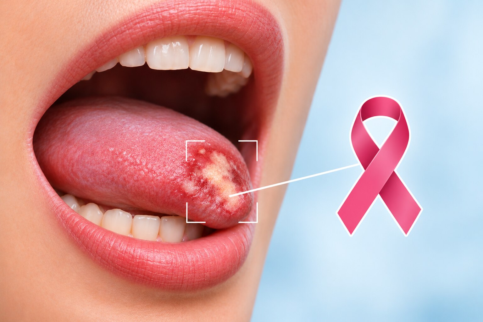
A mouth ulcer lasting more than two weeks, a red or white patch, an oral lump, or a thickened area in the mouth needs examination; the time threshold signals the need for assessment, not a cancer diagnosis.
Other warning signs include:
- Unexplained bleeding, persistent pain, numbness, or tenderness in the tongue or mouth
- A loose tooth without a clear dental cause
- Difficulty chewing, swallowing, moving the tongue, or opening the jaw
- A persistent sore throat, voice change, or lump in the neck
| Situation | What it can indicate | Action |
|---|---|---|
| Breathing difficulty, inability to swallow saliva, rapidly enlarging neck swelling, or heavy bleeding | A potentially urgent airway or bleeding problem | Seek emergency care now |
| Persistent ulcer, red-and-white change, oral lump, neck lump, numbness, or unexplained bleeding | A lesion or symptom requiring direct examination | Book an urgent appointment within days |
| A sore area after biting, a sharp tooth, or denture rubbing that improves and disappears within two weeks | Local irritation that is healing | Arrange routine dental review if it recurs or fails to clear |
Do not wait for pain: early concerning lesions can be painless. A sharp tooth or denture may explain an ulcer, but the area still needs review when it remains after the irritation is corrected or lasts beyond two weeks.
How to Check Your Mouth and Separate Irritation from a Persistent Lesion
Use a bright light and mirror for an oral self-examination, but do not treat a normal-looking mouth as proof that nothing is wrong. A lesion under the tongue or on the floor of the mouth is easy to miss, and early disease can be painless.
- Remove dentures, mouthguards, or dental appliances and wash your hands.
- Look at your lips, gums, and the inside of each cheek. Check for an ulcer, red or white patch, lump, thickened area, bleeding, or colour change.
- Open your mouth wide and inspect the roof, back of the mouth, and both sides of the tongue.
- Gently lift your tongue to examine its underside and the floor of your mouth. Use clean fingers to feel for a firm area, swelling, or tenderness.
- Note its location, size, and appearance. Photographing it beside a ruler helps you detect change.
A sharp tooth ulcer or denture irritation should start improving after the sharp edge is smoothed or the denture is adjusted. Arrange professional assessment if the area remains after two weeks, enlarges, changes colour, becomes firmer, or returns after seeming to heal.
Do not wait for pain. A painless oral lesion, persistent lump, or thickened patch still needs examination. A dentist or clinician must assess any nonhealing area, including one blamed on tooth trauma, habitual biting, or a denture.
Which Risk Factors and Misconceptions Should You Discuss?
The link between smoking and oral cancer is strong, but cigarettes are not the only concern. Tell a clinician about every tobacco product, alcohol use, previous mouth lesions, and past head-and-neck cancer treatment.
Risk increases when tobacco and alcohol are used together. Chewing products also matter: gutkha and khaini contain tobacco, while areca nut or supari can damage oral tissue even without tobacco.
| Exposure | Why it increases concern | What to disclose |
|---|---|---|
| Smoking | Tobacco chemicals injure cells and promote cancerous change | Cigarettes, bidis, cigars, or vaping history |
| Gutkha or khaini | Tobacco exposure remains harmful even when you do not smoke | Product, frequency, and years of use |
| Areca nut or supari | Areca nut is carcinogenic and can cause progressive mouth stiffness | Plain supari, betel quid, or gutkha use |
| Alcohol | Heavy use irritates tissue; combined use with tobacco raises risk further | Type, amount, and frequency |
| Previous disease | Earlier lesions or head-and-neck cancer treatment can signal higher risk | Biopsy results, surgery, radiation, or chemotherapy |
A common misconception is that only older smokers develop concerning lesions. HPV, especially HPV-16, is linked to cancers of the tonsil and base of the tongue, including in people who never smoke. A normal-looking mouth does not exclude an HPV-related throat cancer when a neck lump, swallowing difficulty, or one-sided throat or ear symptom persists.
Pain is not a safety test. A painless patch, lump, thickened area, or ulcer still needs assessment if it remains for more than two weeks.
What Does a Dentist or Clinician Do During Evaluation?
A clinician begins by asking how long the change has been present, whether it is growing, bleeding, painful or numb, and whether chewing, swallowing, speaking or moving the tongue has changed. They also review tobacco, gutkha, khaini, supari, alcohol use, previous oral lesions and earlier head-and-neck cancer treatment.
- A dental assessment checks the lips, gums, cheeks, palate, tongue, floor of the mouth and the area behind the last molars. The clinician lifts the tongue and examines its sides and underside, where a lesion can hide from a mirror.
- An oral examination uses both sight and touch. The clinician feels for firmness, thickness, tenderness, fixation and bleeding instead of judging a patch or ulcer by colour alone.
- A neck examination checks for enlarged or hard lymph nodes, their location, mobility and tenderness. The clinician may also assess jaw movement, tongue movement and sensation.
Appearance cannot reliably separate irritation, a precancerous change and cancer. A painless lesion still deserves assessment, and a normal-looking surface does not rule out disease when symptoms persist.
If the area remains suspicious, the clinician removes tissue for laboratory analysis. An incisional biopsy takes a portion of a larger lesion; an excisional biopsy removes a small, clearly defined lesion. A pathologist examines the sample under a microscope, because histopathology—not appearance, a routine visual check or a commercial screening adjunct—provides the tissue diagnosis.
What Happens After a Biopsy or Abnormal Screening Result?
An abnormal oral screening result is a prompt for diagnostic assessment, not a cancer diagnosis. Visual inspection and commercial screening aids cannot reliably separate irritation, precancerous change, and cancer; a persistent or suspicious lesion usually needs an incisional or excisional biopsy.
1. Ask what the tissue shows. The histopathology report may describe benign inflammation, dysplasia, carcinoma in situ, or invasive cancer. It may also state whether the sample is adequate and whether cancer reaches the specimen’s edges, called margins.
2. Confirm the next step with the clinician who arranged the test. Benign findings may need treatment of a sharp tooth, denture trauma, infection, or observation with a defined review date. Dysplasia or cancer requires specialist planning rather than indefinite monitoring.
3. Use oral cancer scans when the report or examination indicates possible cancer. Imaging such as ultrasound, contrast-enhanced CT, or MRI helps assess the tumour’s depth, nearby tissues, lymph nodes, and distant spread; scans do not replace biopsy.
Specialist input is useful when the lesion persists despite a reassuring result, the biopsy sample is small or unclear, dysplasia is reported, lymph nodes or a neck lump are present, or treatment would affect speech, swallowing, or appearance.
A second opinion from a head-and-neck or surgical oncology team can clarify conflicting reports and compare surgery, surveillance, or further testing. Dr.Parag Ingle - Surgical Oncologist can help interpret the report alongside examination and imaging when the diagnosis or next step remains uncertain.
Related services
Oral cancer treatment involves various medical procedures to address cancerous growth in the mouth. View service → |
Cancer screening Our Cancer Screening service is a vital health check aimed at detecting cancer in its early stages, ensuring timely intervention for better outcomes. View service → |
Frequently asked questions
Which mouth and neck changes need prompt attention?
Arrange a dental or medical appointment for a sore, ulcer, lump, thickened area, red patch, white patch, bleeding site, numbness, loose tooth, swallowing difficulty, voice change, or neck lump that lasts beyond two weeks. Seek urgent care for breathing difficulty or rapidly increasing swelling.
How can I check my mouth and separate irritation from a persistent lesion?
Use a bright light and a mirror to inspect your lips, gums, inner cheeks, tongue, floor of the mouth, and palate. Note the location, size, colour, pain, bleeding, and duration. Remove an obvious irritant when possible, but arrange an examination if the change remains after two weeks or worsens sooner.
Which oral cancer risk factors and misconceptions should I discuss?
Discuss tobacco in any form, areca nut or gutkha, alcohol use, prior oral lesions, human papillomavirus exposure, and previous head and neck cancer. Do not dismiss a lesion because it is painless, because you are young, or because you do not use tobacco.
What does a dentist or clinician do during an oral cancer evaluation?
The clinician asks when the change began, examines the mouth and throat, feels the tissues and neck for lumps, and checks nearby lymph nodes. They may photograph or measure the area and refer you for specialist assessment or biopsy.
What happens after a biopsy or abnormal oral cancer screening result?
A screening result identifies an area that needs assessment; it does not confirm cancer. A biopsy examines tissue under a microscope. Your clinician explains the pathology result and, if cancer is found, may arrange imaging, staging tests, and a treatment discussion with the appropriate specialists.
Related service
Cancer is a disease that causes abnormal growth of cells in the body. It can affect any part of the body and can spread to other organs. View service → |